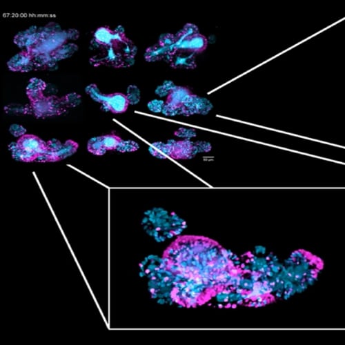
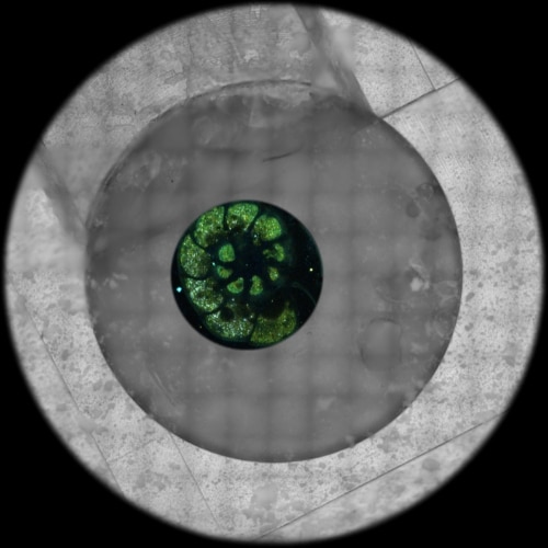
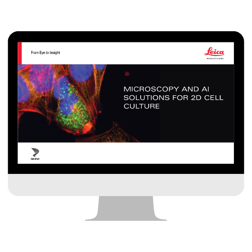
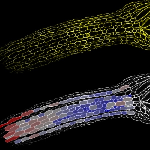
ON-DEMAND EVENT See the Hidden at Micron | Visualizing Organoid Complexity Through Advanced Imaging WATCH NOW MASTERCLASS How to Choose the Right Confocal Microscope for Your Research Register for free expert insights, comparison guides, and technical resources delivered weekly to your inbox. REGISTER NOW Previous Next Advanced Imaging of Organoid ModelseBookStreamlined High Pressure Freezing for Cryo-ET: Waffle Method to Serial Lift-OutWebinarIlluminating Nanomaterial–Cell Interactions by Confocal MicroscopyWebinarChoosing Your Next Confocal: What Experts Look For—and Why It MattersWebinarDemocratizing Nanoscale Imaging at Molecular ResolutionWebinarAdvancing Precision and Control in EM Micro Target PreparationWebinarMultiscale Imaging Workflows for Organoid ImagingWebinarSee the Hidden: Advancing Cryo Workflows for Cellular DiscoveryWebinarMicroscopy and AI Solutions for 2D Cell CultureeBookImage Segmentation to Study Plant BiologyWebinarIntegrated Solutions for Serial Sectioning and Cryo-EM with UC EnuityWebinarGet to Insights Faster and Easier with AI Image Analysis ToolsWebinarUnlocking the Secrets of Organoid Models in Biomedical ResearchWebinarSee the Hidden: Discover More with FLIMWebinarOvercoming the High Multiplexing Barrier: 3D Spatial Omics in One GoWebinarDesigning the Future: Novel, Scalable Stem Cell CultureWebinarFrom Bench to Beam: A Complete Correlative Cryo Light Microscopy WorkflowWebinarAI-Powered 3D Organoid AnalysisWebinarMulti-View Light Sheet Microscopy to Track Cell Lineages in Tissue MorphogenesisWebinarEnhancing 3D Spatial Biology with AI: Simplified Insights for AllWebinarPrecision Sectioning Redefined: Bringing Ultramicrotomy to the Digital AgeWebinarImaging GPCR Activity: Unravelling Mechanisms from Molecules to MorphologyWebinarNew TauSTED Tools for Gentle Live Imaging at Nanoscale ResolutionWebinarCellular Crosstalk in Neurodevelopmental DisordersWebinarMultiplex Imaging to Visualize Liver Cancer Stem Cell NichesWebinarConsiderations for Multiplex Live Cell ImagingeBookThe Power of Spatial Biology: A Microscopy GuideeBookMicroscope Cameras for Life ScienceWhitepaperWhich Stereo Microscope Is Right for You?WhitepaperElevate Your Research with Spatial InsightsWebinarA Platform for Exploring Subcellular Spatial PhenotypesWebinarSee the Hidden: Adding Time to the Correlative EquationWebinarReveal Hidden Information in Your Microscopy Experiments With the Power of AIWorkshopA Novel Human Brain Organoid Model to Study Microglia Phenotypes in Health and DiseaseWebinarWindows On Neurovascular PathologiesWebinarAccelerate Scientific Progress: The Power of Reproducibility, Collaboration and New Imaging TechnologiesWebinarBreaking the Ice: Freeze Fracture for Cryo SEMWebinarSee the Hidden: Spatial ProteomicsWorkshopVirtual Reality Showcase for Stellaris: Leica’s Confocal Microscopy PlatformWebinarMultiplex Annotated Tissue Imaging System (MANTIS®)WebinarSee the Hidden: Correlative Techniques for Electron MicroscopyWorkshopAdvanced Cryo Preparation TechniquesWebinarFive-Color Sted With a Single Depletion Laser and Fluorescence Lifetime Phasor SeparationWebinarThe Role of Iron Metabolism in Cancer ProgressionWebinarUltramicrotomy:The Cutting Edge of SectioningWebinarSee the Hidden: Spatial Biology IIWorkshopLive-Cell Fluorescence Lifetime Multiplexing Using Organic FluorophoresWebinarSee the Hidden: Translational Cancer ResearchWorkshopSee the Hidden: Spatial BiologyWebinarVisualizing Protein-Protein Interactions by Non-fitting and Easy FRET-FLIM ApproachesWebinarFree eBook: Microscopy for Neuroscience ResearcheBookDiscover a New Era in Microscopy. Meet Mica.WebinarLive Imaging Across Scales and TimeeBookCoral Cryo: Access What MattersWebinarLAS X Navigator Tutorial Navigating Live Cell ImagingWebinarThe POWER of STELLARIS…WebinarThe POTENTIAL of STELLARIS…WebinarThe PRODUCTIVITY of STELLARIS…WebinarAdding Dimensions to Multiplex Molecular ImagingWebinarRegulators of Actin Cytoskeletal Regulation and Cell Migration in Human NK CellsWebinarDissecting Proteomic Heterogeneity of the Tumor MicroenvironmentWebinarOptimizing THUNDER Platform for High-Content Slide ScanningWebinarHigh-Pressure Freezing: Revealing Functional Mechanisms of Synaptic TransmissionWebinarRevealing Cellular Dynamics with Millisecond Precision – The New Tool That Turned Electron Micrographs into Motion Pictures of Neural CommunicationWebinarHow to Conduct Localized Proteomics of Microscopic RegionsWebinarOptimized Cryo Workflows Using the Vacuum Cryo Transfer System VCT500WebinarAdvances in Cellular Imaging by Cryo-3D Correlative Light and Electron TomographyWebinarLive Cell Isolation by Laser MicrodissectionWebinarExpanding the Limits of Electron Microscopy Sample Preparation with the Leica EM ICE High Pressure FreezerWebinarArray Tomography for SEM 3D ReconstructionWebinarInvestigating Synapses in Brain Slices With Enhanced Functional Electron MicroscopyArticleRevealing Functional Mechanisms of Synaptic Transmission by High-Pressure FreezingWebinar