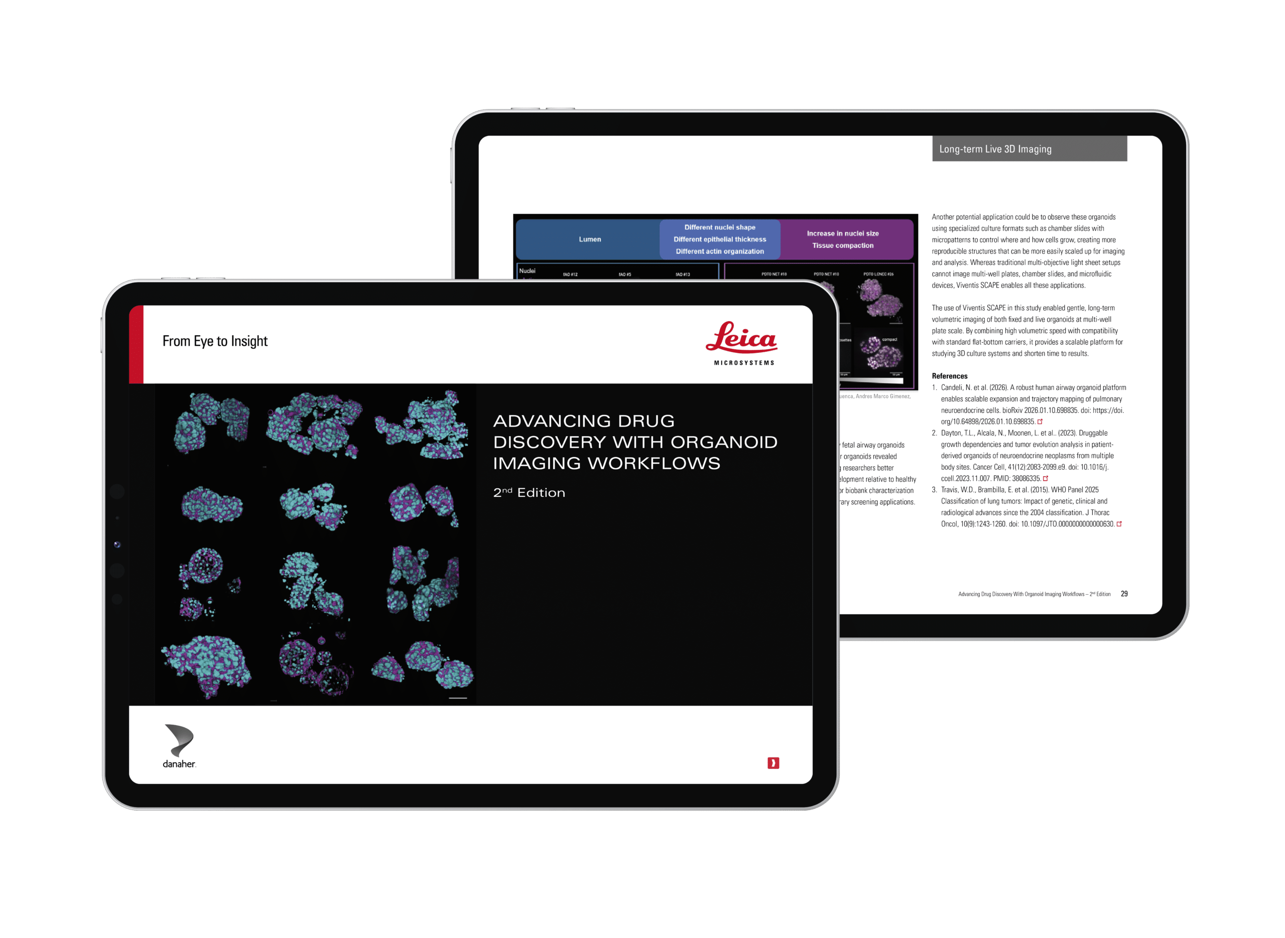
Advanced Imaging of Organoid Models
Advancing drug discovery with imaging solutions for complex human-relevant in vitro models
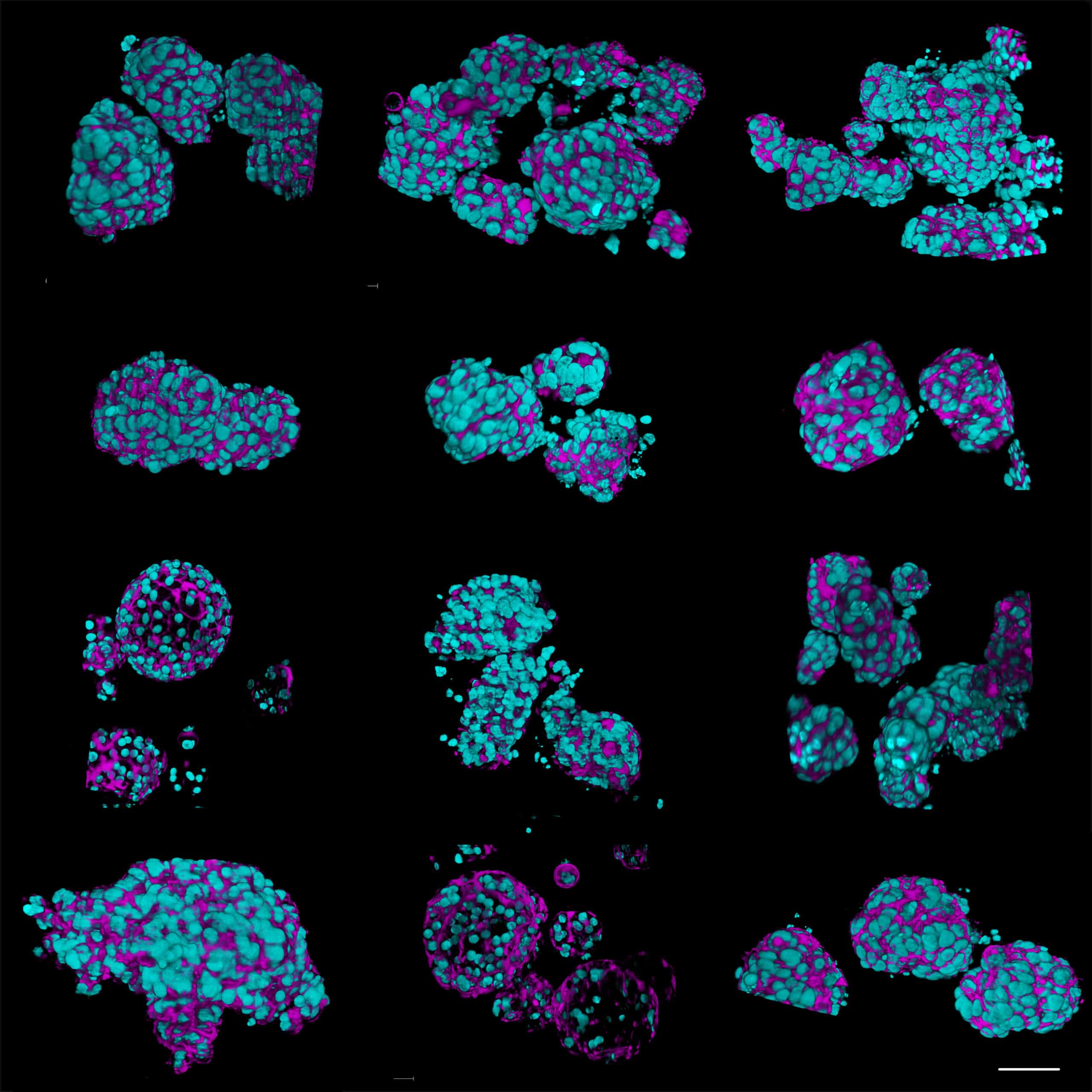
Complex In Vitro Models (CIVMs) such as organoids are becoming increasingly important in early drug discovery and translational research, supporting the shift toward more predictive, human-relevant data in line with evolving regulatory expectations.
However, extracting meaningful information from these models is not straightforward. Their size, heterogeneity and scalability, combined with sensitivity to handling and photodamage—can make robust imaging and characterization challenging.
The second edition of this popular eBook provides a practical, workflow-oriented guide to organoid imaging and analysis. It is supported by real-world examples from biotech and translational research.
Key learnings
-
Identify the right imaging approach for your organoid application —and learn how complementary techniques work together
-
Understand key challenges when imaging dense 3D multicellular systems and how to overcome them
-
Capture dynamic organoid biology over time with gentle, long-term live imaging approaches
-
Scale experiments to higher-throughput workflows using multi-well plate-compatible imaging
-
Learn from real-life case studies including disease modeling, drug uptake, regenerative medicine, and biobank characterization