From Bench to Beam: A Complete Correlative Cryo Light Microscopy Workflow
How correlative light electron microscopy can empower structural biology research
Dr. Andreia Pinto
Advanced Workflow Specialist, Leica Microsystems
Read BioDr Pinto started her career as a Biomedical Scientist in Histocellular Pathology with a particular interest in electron microscopy. In 2014, she worked at the Primary Ciliary Diagnosis (PCD) in Lisbon. In 2019, Andreia moved to the Royal Brompton Hospital, London, working as a Thoracic Research Associate, where she was responsible for training a deep machine learning platform to recognize patterns in EM images of cilia in diagnosing PCD. She completed her PhD on this topic in 2022 while also investigating new insights into SARS-CoV-2 infection of the respiratory airway. Andreia is currently an Advanced Workflow Application Specialist with Leica Microsystems, based at the Imaging Centre of the European Molecular Biology Laboratory (EMBL) in Heidelberg.
Close
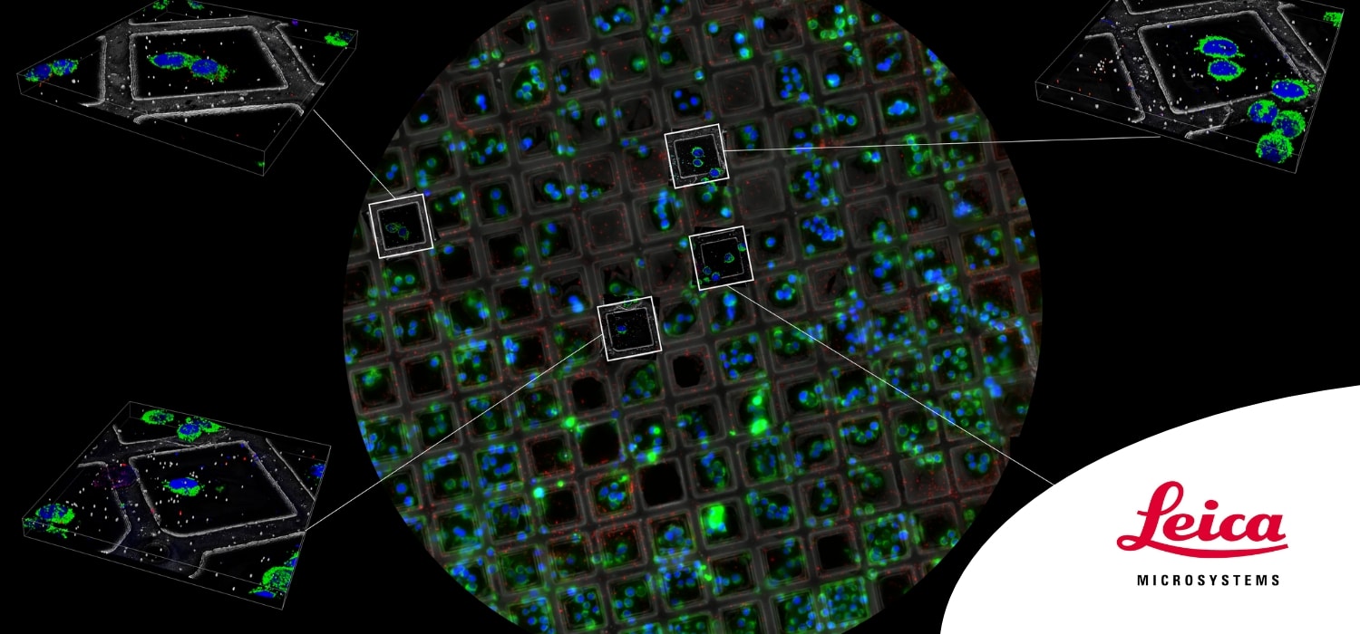
Correlative imaging techniques are at the heart of the crusade for better understanding of subcellular biological structures. The use of Correlative Workflows, including Cryo EM, Sub-tomogram averaging, Cryo-FIB, and 3D Volume Imaging, are powerful tools in the field of (in-situ) structural biology, allowing researchers to obtain higher and higher resolution images of complex biological specimens such as organoids and 3D cultures.
In this webinar, experts from the European Molecular Biology Laboratory and Leica Microsystems explore advanced techniques that comprise Correlative Workflows. They cover the full workflow: from the best sample preparation practices, through FIB milling and Electron Microscopy techniques, to image processing and 3D reconstruction.
Key Learnings:
- Understanding the techniques required for Correlative Workflows with Cryo FIB and Volume EM, from the workbench to electron beam
- Overview of the use of correlative techniques such as Cryo EM, sub-tomogram averaging and 3D Volume Imaging for structural biology research
- Application examples of how these techniques have been used to advance our understanding of biological structures
