Adding Dimensions to Multiplex Molecular Imaging

Professor Scott Fraser
University of Southern California, Departments of Molecular and Computational Biology, of Biomedical Engineering and of Stem Cell Biology and Regenerative Medicine, Los Angeles, CA
Molecular imaging of living specimens offers a means to draw upon the growing body of high-throughput molecular data to better understand the underlying cellular and molecular mechanisms of complex events ranging from embryonic development to disease processes. However, imaging approaches are challenged by unavoidable trade-offs between spatial resolution, temporal resolution, field of view and the limited photon budget.
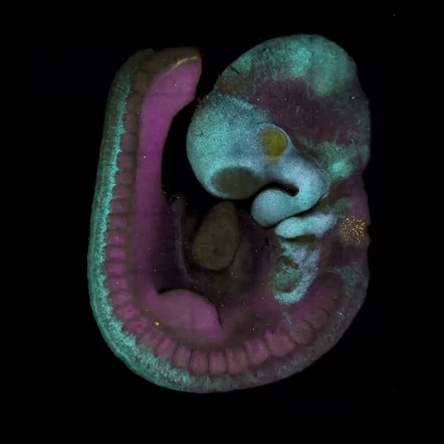
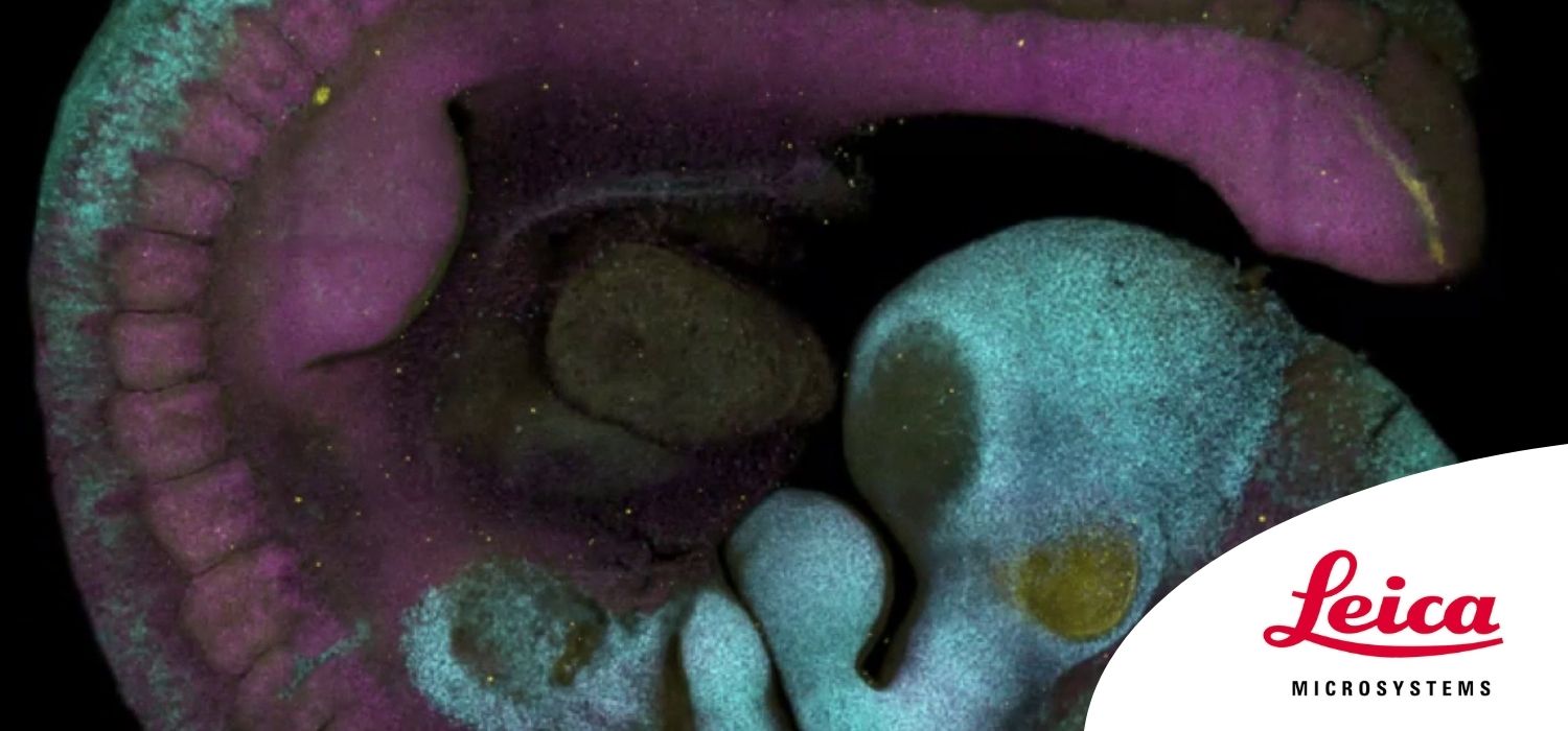
We are attempting to advance this trade-off by constructing faster and more efficient light sheet microscopes that maintain subcellular resolution. Our two-photon light-sheet microscope combines the deep penetration of two-photon microscopy and the speed of light sheet microscopy to generate images with more than ten-fold improved imaging speed and sensitivity. This combination of attributes permits 4D cell and molecular imaging with sufficient speed and resolution to generate unambiguous tracing of cells and signals in intact systems.
To increase the 5th Dimension, the number of simultaneous labels, we are refining new multispectral image analysis tools that exceed the performance of our previous work on Linear Unmixing by orders of magnitude in speed, error propagation and accuracy. These new analysis tools permit rapid and unambiguous analyses of multiplex-labeled specimens.
In parallel, we have refined label-free approaches so that imaging and sensing can be extended to patient-derived tissues and even human subjects. The low concentrations and low sensitivity of the techniques can make single cell approaches challenging. We are refining fluorescence lifetime approaches (FLIM), combining it with multispectral tools to optimize intravital imaging in these challenging settings.
Combined, these imaging and analysis tools offer the multi-dimensional imaging required to follow key events in intact systems and allow us to use noise and variance as experimental tools rather than experimental limitations.
Learning Objectives:
- Biological imaging involves trade-offs between resolution, speed, depth, and field of view
- Multiplex tools can increase the efficiency and the number of labels that can be imaged
- Fluorescence lifetime is a powerful but underused tool for intrinsic and extrinsic labels
- Phasor approaches can offer more efficient processing, especially in low light conditions
Originally posted by Leica Microsystems here.