See the Hidden: Spatial Biology
Dr Abdullah Ahmed
Advanced Workflow Specialist Leica Microsystems
Read BioAbdullah joined Leica Microsystems in 2019 as an Advanced Workflow Specialist. Before this, he pursued a joint Ph.D. project with Oxford Brookes University and Evotec, focusing on the characterization of the mechanisms of cancer signaling to provide novel targets and cures. Based at the Central Laser Facility (Oxford), his research focused on mTOR signaling by employing fixed-cell and live-cell imaging using confocal microscopy. He developed and used fluorescent imaging technologies to observe the localization and interactions of proteins in conjunction with FRET–FLIM (Förster Resonance Energy Transfer measured by Fluorescence Lifetime Imaging Microscopy). Furthermore, super-resolution microscopy techniques, important to aspects of the research project and for driving the bio-imaging facility at OCTOPUS (STFC), were imperative during his doctorate.
Close
Ronald N. Germain, M.D., Ph. D.
Chief, Laboratory of Immune System Biology and Lymphocyte Biology Section, NIAID (NIH)
Read BioRonald received his M.D. and Ph.D. from Harvard University in 1976 and is currently Chief of the Laboratory of Immune System Biology at the National Institute of Allergy and Infectious Diseases, NIH. Over the years, he and his colleagues have made vital contributions to our understanding of Major Histocompatibility Complex class II molecule structure–function relationships, the cell biology of antigen processing, and the molecular basis of T cell recognition. More recently, his laboratory has focused on the relationship between immune tissue organization and control of adaptive immunity studied by utilizing novel dynamic and static in situ microscopic methods that his laboratory helped pioneer. Dr. Germain has published over 400 scholarly research papers and reviews and trained more than 70 postdoctoral fellows. Several of them hold senior academic and administrative positions at leading universities and medical schools worldwide.
Close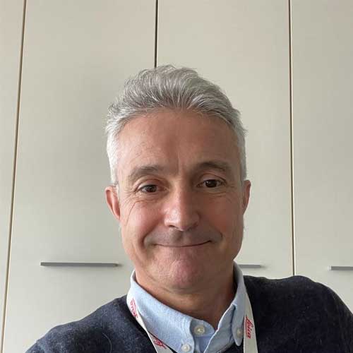
Dr. Mauro Luca Baron
Advanced Workflow Specialist Leica Microsystems
Read BioMauro received his degree in Biology from the University of Milan, where he focused on Human Pathology. He then worked for two years in the Institute of Internal Medicine at the University of Milan on innovative techniques for diagnosing Helicobacter pylori and HCV. After his scientific career, he moved to a company involved with diagnostic products, where he spent five years as a Product Specialist in Italy. In 2000, he joined Leica Microsystems Italy as Product Manager Microscopy, covering all aspects of microscopy applications – particularly those related to Laser Microdissection (LMD). In 2009, he became European Field Support Specialist for the Life Science Research Division, primarily responsible for microdissection-related topics in EMEA. Since 2019, Mauro has provided application support for the THUNDER widefield imaging systems in Italy and, more recently, workflows for the Cell DIVE and Mica systems and their applications in multiplexing. He has presented at scientific conferences and hosted workshops all over EMEA, and organized remote training activities for researchers. Mauro enjoys interacting with scientists with different expertise and specimens to find the best applications and imaging workflows.
Close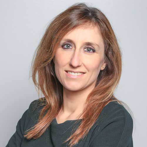
Dr. Berta Cillero-Pastor
Group leader, Department of Cell Biology-Inspired Tissue Engineering MERLN Institute, Maastricht University
Read BioBerta studied molecular biology and biochemistry at the Autonomous University of Madrid, Spain. She obtained her Ph.D. at the INIBIC Institute of La Coruña (cum laude), having been awarded a fellowship from the Carlos III Health Institute (Spain) to study the effect of pro-inflammatory cytokines in diseased cartilage. After obtaining an Angeles Alvariño fellowship, she moved to Amsterdam to work as a postdoctoral researcher at AMOLF. Here, she developed new mass spectrometry imaging approaches in orthopedics. In 2015, she joined Maastricht University as CORE lab leader in the division of Imaging Mass Spectrometry (M4i). She has since established her own research line applying mass spectrometry imaging and proteomics for different biomedical applications, focusing on cardiovascular research and musculoskeletal diseases. As principal investigator at MERLN, she develops and applies spatial omics approaches to understand cell-biomaterial interactions, local drug delivery, biofilm formation, and molecular mechanisms of cell differentiation.
Close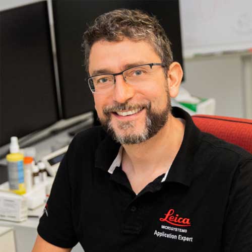
Dr. Jens Peter Gabriel
Advanced Workflow Specialist Leica Microsystems
Read BioJens obtained his Ph.D. in Biology (Neurobiology) from the University of Cologne. He continued to work in this field as a postdoctoral researcher at the Max-Planck Institute for Medical Research (Heidelberg) and the Karolinska Institute (Stockholm). His primary interest was neuronal networks generating behavior, which he studied in zebrafish using methods like electrophysiology, multi-photon calcium imaging, and confocal microscopy. His passion for biological imaging was why Jens joined Leica Microsystems in 2009. As an Advanced Workflow Specialist he is now contributing to scientific progress in different ways: by helping researchers identify which microscope system best fits their requirements and by supporting them through training and application advice.
Close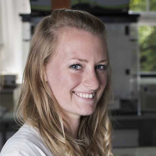
Dr. Lindsey Marshall
EMEA Customer Success Manager for Aivia AI Image Analysis Software Leica Microsystems
Read BioLindsey obtained her Ph.D. in Physiology and Molecular Biology at the National Museum of Natural History (France), followed by post-doctoral research in Regenerative Medicine at the University of Manchester (UK). She began working with state-of-the-art software at a start-up specializing in the application of virtual reality for education and exploration of large microscopy data stacks. Seeing Artificial Intelligence (AI) as the key to significantly speeding up microscopy image analysis, she is delighted to now work with Aivia software. Lindsey enjoys interacting with scientists and especially helping non-programmers to easily apply AI to their analysis workflow, ultimately helping them obtain outstanding results with their images.
CloseIn this next highly anticipated virtual edition of our See the Hidden Workshop Series, we will start to examine some of the emerging microscopy techniques being used to study cells within a spatial 2D and 3D context. We will examine the behavior and identity of cells in diverse tissues, and present proof-of-concept studies to identify local changes in protein content and investigate pathways involved in cardiac tissue injury. New technologies and approaches that leverage fluorescence multiplexing and spatial omics to address spatial cell biology will be discussed, along with the molecular insights they can bring to the treatment of diseases such as heart disease, cancer and immunology.
This online event, featuring live panelists, brings together a series of scientific talks and microscopy showcases. Dr. Ronald N. Germain (NIH) and Dr. Berta Cillero-Pastor (Maastricht University) will talk about how specialized microscopy approaches contribute to their spatial biology research. You will discover streamlined workflows that can enable a better understanding of biological pathways and provide greater knowledge for translational research.
The program centers around four specific areas in microscopy:
- widefield multiplexing
- single-cell/tissue microdissection
- high-resolution confocal microscopy
- Artificial Intelligence (AI)-based analysis.
We will continue to address the theme of advanced microscopy techniques for spatial biology with a follow-up workshop later in the year – so don’t miss this opportunity to join us for part 1!
Welcome and introductions
Dr. Abdullah Ahmed
Panel discussion on emerging microscopy trends in spatial biology
Gaining insight into tissue biology using highly multiplex 2D and 3D tissue imaging
Dr. Ronald Germain
Obtaining spatial context with clarity using widefield microscopy
Dr. Mauro Baron Luca
Spatial omics and beyond
Dr. Berta Cillero-Pastor
Get closer to the (spatial) truth with the STELLARIS Confocal Microscope Platform
Dr. Jens Peter Gabriel
Aivia – the future of AI microscopy
Dr. Lindsey Marshall
Closing remarks
Dr. Abdullah Ahmed
