Array Tomography for SEM 3D Reconstruction

Frank Assen
Read BioFrank Assen has a bachelor’s degree in Technical Medicine and a master’s degree in Reconstructive Medicine from the University of Twente (twenty), The Netherlands. He is currently a doctoral student in the group of Michael Sixt at IST Austria in Vienna, Austria, where he is working on the role of stromal cells in lymph node swelling during inflammation.
Close
Robert Ranner
Read BioRobert Ranner, is Product Manager EM Specimen Preparation at Leica Microsystems, Nanotechnology Division based in Vienna. He completed his education in optics and mechatronics before joining Leica Microsystems GmbH in 1985. As product manager Robert is responsible for ultramicrotomy, ion etching and solid state preparation instruments. Robert has fifteen years of application experience in industrial sample preparation for transmission electron microscopy (TEM) and scanning electron microscopy (SEM).
CloseTutorial abstract
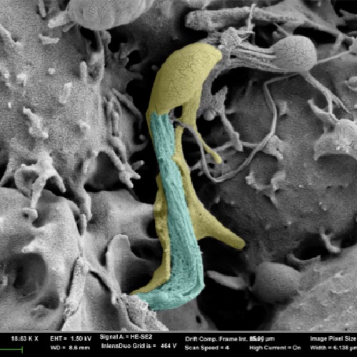
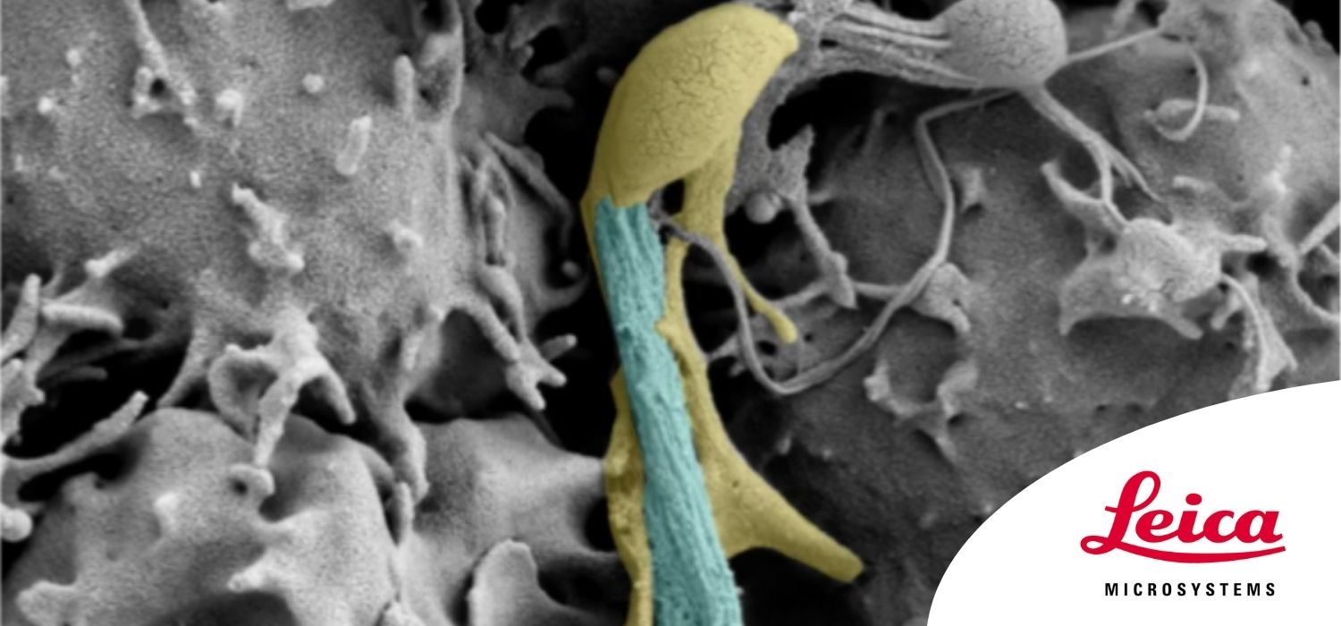
Array tomography (AT) is a 3D image reconstruction technique for high resolution, quantitative analysis of biological structures. For optimal results, ultrathin and ordered sections are an absolute requirement.
In this webinar you will get tips and tricks to optimize the workflow of your array tomography:
- Fast and precise trimming of the sample block-face
- Adhesion of a single section to create ribbons
- Automated serial sectioning with the ARTOS 3D ultramicrotome
- Acquisition and processing of a 3D SEM dataset
- Segmentation and 3D reconstruction of cells
- Interpretation of 3D reconstructions