A Novel Human Brain Organoid Model to Study Microglia Phenotypes in Health and Disease

Simon T. Schäfer
Assistant Professor, Technical University of Munich (TUM)
Read BioProfessor Simon T. Schäfer studied medicine and biomedical sciences in Greifswald and San Diego and received his PhD in 2015. He then joined the Salk Institute for Biological Studies as a postdoctoral fellow, before joining the Department of Psychiatry at TUM as an Assistant Professor for advanced organoid technologies. His lab is part of the newly-established Center for Organoid Systems. His current research focuses on advancing novel stem cell-based technologies to generate three-dimensional models that recapitulate the structural and functional organization of the human brain.
CloseSee how researchers at the Technical University of Munich are advancing novel stem cell-based technologies in order to generate three-dimensional (3D) models that recapitulate the structural and functional organization of the human brain. These technologies are being used to push the boundaries for personalized research on human-specific brain disorders and to identify strategies for facilitating brain repair.
In this webinar, you will learn:
- How novel organoid models enable research into areas that were historically difficult to study
- The different types of functional human microglia phenotypes and their impact on brain health
- Techniques to model interactions between the human brain environment and microglia
Microglia are specialized brain-resident immune cells that play crucial roles in brain development, homeostasis, and disease. However, until now, the ability to model interactions between the human brain environment and microglia has been severely limited.
In this webinar, Professor Simon T. Schäfer will present how a novel organoid model enables his group to investigate functional human microglia phenotypes under physiological and pathological conditions—which is a critical next step for modeling human brain environment-dependent microglia phenotypes in health and disease.
Many of the images that will be presented in this webinar were acquired using the THUNDER Imager Live Cell & 3D Assay from Leica Microsystems.
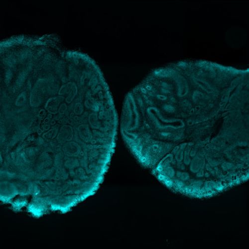
Banner image: Brain organoid section (stained with DAPI) acquired using the Leica THUNDER Imager. Image courtesy of Janina Kaspar and Irene Santisteban, Schäfer Lab, TUM.
Photo credit: Technical University of Munich (TUM).