Live Cell Isolation by Laser Microdissection

Dr. Oleg Podgorny
Read BioDr. Oleg Podgorny is a senior researcher at Koltzov Institute of Developmental Biology of Russian Academy of Sciences and Federal Research and Clinical Center of Physical-Chemical Medicine, Moscow, Russia. He received his Ph.D. at Koltzov Institute of Developmental Biology in 2006. After Ph.D. thesis defense, being focused on stem cell maintenance in vitro, he acquired experience on the use of laser microdissection for live cell isolation. In 2015, he joined professor Grigori Enikolopov’s group at Stony Brook University, New York USA, as a postdoctoral associate. His research was focused on elucidating modes of neural stem cell division and understanding mechanisms underlying their maintenance in the adult brain. This year, Dr. Oleg Podgorny returned in Moscow and joined the teams at Koltzov Institute of Developmental Biology
CloseIn this tutorial, you will learn:
- How to best prepare your specimen for live cell isolation by laser microdissection
- How to optimize your laser microdissection workflow
- How to avoid common pitfalls of this technique
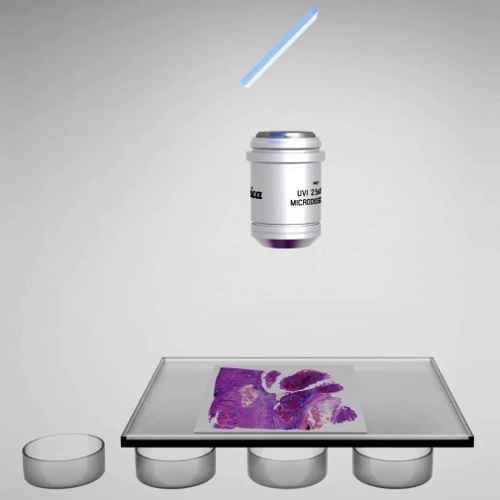
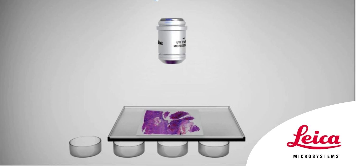
Laser microdissection is a tool for the isolation of homogenous cell populations from their native niches in tissues to downstream molecular assays. Beside its routine use for fixed tissue sections, laser microdissection may be applied for live cell isolation. Unlike other well-established and widely used techniques for live cell isolation and single cell cloning—such as FACS, MACS, cloning by limited dilution, and so on—laser microdissection allows for capturing live cells and cell colonies without their detachment from the carrier. In other words, there is no need to prepare a single cell suspension before the isolation procedure using mechanical and enzymatic dissociation, which can affect cell fate after plating. This feature of laser microdissection is desirable for stem cell research. We established a simple strategy for the efficient live cell isolation using the Leica Laser Microdissection platform. We were able to demonstrate not only colony formation from the isolated samples containing live cells, but also single cell cloning. In this webinar, specimen preparation, laser adjustment, overall workflow, and limitations on live cell isolation by laser microdissection are discussed.