Unlocking the Secrets of Organoid Models in Biomedical Research
Dr Tanya Singh
Postdoctoral Research Scientist, Salman and Wade-Martins Lab, Department of Physiology, Anatomy and Genetics, University of Oxford
Read BioDr. Tanya Singh began her research at NBRC, India, focusing on iPSC reprogramming and miRNA-regulated gene expression in stem cells. During her PhD at Cardiff University, she studied the impact of copy number variations on glial cells in neuropsychiatric disorders. Her postdoctoral work at Oxford University led her to study epileptogenesis using 2D and 3D stem cell models of Tuberous Sclerosis. Recently, working with the Wade-Martins group, she has focused on Parkinson’s disease, generating ventral mid-brain organoids and Enteric Nervous System (Gut-Brain axis) using stem cells and microfluidics.
Close
Dr Ramon Garcia Maset
Research Fellow, Rohn Group, University College London (UCL)
Read BioDr. Ramon Garcia Maset, a Research Fellow in Professor Jennifer Rohn's lab at UCL, currently works at the University of Oxford in Professor Dario Carugo's lab. He is part of the EPSRC-funded "Beyond Antibiotics" project, focusing on urinary tract infections (UTIs), 3D microtissue modeling, host-pathogen interactions, biofilms, and antimicrobial resistance. Dr. Garcia Maset earned his PhD from the University of Warwick, working with the Perrier and Harrison groups on antimicrobial and antibiofilm polymers to address wound and respiratory infections.
Close
Dr Annalisa Rizza
Advanced Workflow Specialist, Widefield Microscopy, Leica Microsystems
Read BioDr. Annalisa Rizza currently works as an Advanced Workflow Specialist in Widefield Microscopy at Leica Microsystems. Prior to this role, she served as a Research Associate at the Sainsbury Laboratory, University of Cambridge (UK), where her research was focused on FRET biosensors. She earned her PhD in Biology from the University of Freiburg (Germany).
Close
When THUNDER Meets Spinning Disk
Join us as we delve deeper into the world of organoids and 3D models, which are essential tools for advancing our understanding of human health.
Navigating these complex structures and obtaining clear images for analysis can be challenging.
In this event, researchers from The University of Oxford and University College London (UCL) join Leica Microsystems scientists to show how combining THUNDER technology with the CrestOptics CICERO spinning disk confocal can provide more convincing, high-quality data for deeper insights into a variety of models.
This event was hosted as part of the ongoing collaboration with the Leica Centre of Excellence at the Micron Bioimaging Facility, University of Oxford
What You’ll Learn:
1. Examining 3D human bladder microtissue infected with E. coli
Jennifer Rohn’s lab at UCL has developed a pioneering 3D human urothelium organoid model (3D-UHU) to explore UTI pathology and treatment failures. Discover how clearer insights provided by the new THUNDER Imager Cell Spinning Disk can help them further explore the impact of bacterial filamentation and intracellular bacterial communities on UTI outcomes.
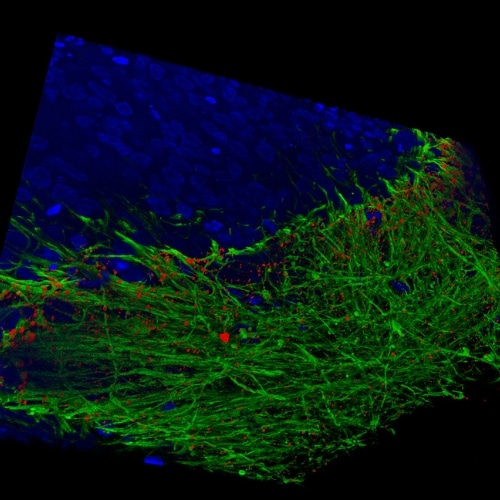
2. Exploring human stem cell-derived mid brain organoids
The Wade-Martins group in Oxford examines disease mechanisms for Parkinson’s disease. See how they develop ventral mid-brain organoids and Enteric Nervous System (Gut-Brain axis) using stem cells and microfluidics and how much more detail they can now uncover.
3. How microtubule dynamics influence cell shape
To illustrate the diversity of samples that can benefit from the synergy of THUNDER and spinning disk, we also share data on individual microtubule dynamics, courtesy of the Sainsbury Laboratory in Cambridge.
This webinar is a must-watch for researchers looking to gain deeper insights into their organoid and 3D models or enhance their live cell imaging capabilities.
About THUNDER Imager Cell Spinning Disk
While spinning disk alone provides fast, versatile, confocal imaging and powerful optical sectioning, the synergistic pairing with THUNDER eliminates residual out-of-focus light, resulting in greater contrast and resolution.
This enables you to extract more statistically significant data from diverse 3D samples in less time and with reduced phototoxicity.
Typical applications include live cells, 3D cell cultures, organoids, spheroids, model organisms, plant biology and thick tissue samples.
Image: Human stem cell-derived mid brain organoids. Courtesy of Dr Tanya Singh, University of Oxford.