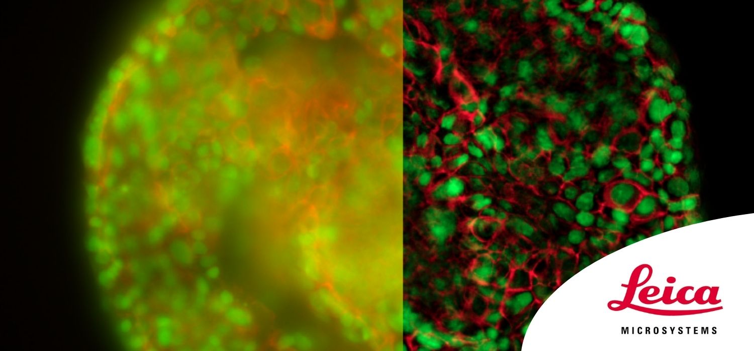
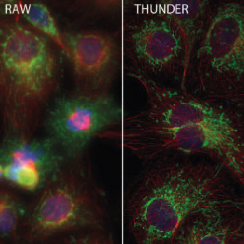
Thunder Imagers: High Performance, Versatility and Ease-Of-Use for Your Everyday Imaging Workflows.


Dr. Falco Krüger
Read BioAdvanced Workflow Specialist Widefield
Life Science Research Division
Falco did his PhD in Biology in the Plant Cell Biology group of Prof. Karin Schumacher at the Centre for Organismal Studies (COS) in Heidelberg, Germany. For his research on vacuole biogenesis in Arabidopsis thaliana, he used several different confocal microscopy techniques to understand the contribution of subcellular compartments to the development of one of the most prominent plant cell organelles.
By joining Leica Microsystems in May 2018, he switched gears, and started focusing on advanced widefield microscopy. Since the THUNDER launch earlier this year he has been hosting many workshops all over Germany, Austria and Switzerland, demonstrating the power of the new THUNDER Imagers to the scientific community. He enjoys getting to know different customers and their specimens, applications and imaging workflows.

Dr. Remco Megens
Read BioRemco Megens is a PI at the Institute for Cardiovascular Prevention (IPEK) of the Ludwig-Maximilians-University in Munich, Germany, and leads its core facility for advanced optical imaging. His major research focus is the development and application of (advanced) microscopic methods and novel labeling strategies for atherosclerosis research. Application of two-photon laser scanning microscopy (TPLSM) for their research has a strong emphasis on its utilization in large arteries and bone marrow. Novel TPLSM methods and labeling strategies that have been developed enable specific imaging of morphological and functional aspects of the target tissues ex vivo and in vivo. The optical nanoscopy modality Stimulated emission depletion (STED) is applied to study atherosclerosis with higher accuracy. Besides utilization of 3D STED (and its core modality Confocal Laser Scanning Microscopy) for in vitro models, they developed applications and staining strategies for multicolor 3D STED in tissues and models specific to the field of cardiovascular research. The IPEK has been working with instant computational clearing (THUNDER) technology to advance both the image quality of standard (immuno)histological samples as well as the overall imaging workflow in the facility.
CloseIn this webinar, you will learn:
- How to get sharp, high quality images from thick 3-dimensional microscope samples
- How to shorten your imaging workflow from image acquisition to data analysis
- A broad spectrum of imaging workflows for a range of life science applications
- Examples of multi-platform imaging using THUNDER Imagers with different systems, such as CLEM, confocal and DLS
Webinar abstract
THUNDER Imagers are a brand-new class of widefield microscopes developed by Leica Microsystems. They can acquire imaging data, even of thick 3-dimensional specimens, with extraordinary quality and speed.
However, there is more to life science experiments than beautiful images. Robust quantification and repeatability of imaging experiments are crucial. THUNDER Imagers provide the full workflow, from image acquisition to data analysis, with an amazing ease of use.
This webinar will showcase the versatility and performance of THUNDER Imagers in many different life science applications: from counting nuclei in retina sections and RNA molecules in cancer tissue sections to monitoring calcium waves in Arabidopsis seedlings and much more.
