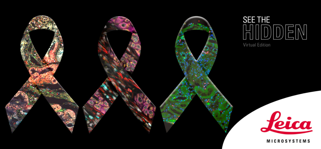
LIVE EVENT
See The Hidden: Virtual Summit on Cancer Research in Microscopy
Life science researchers play a vital role in understanding how and why cancer starts and progresses and developing new diagnostic tools and therapeutic approaches.
We invite you to a special edition of our See the Hidden workshop series. Join us as we dive deep into current developments in cancer research microscopy. You’ll hear from leading researchers about their recent accomplishments in cancer imaging, cancer evolution, biomarker research, and image analysis.
Plus, explore the latest cancer imaging products and solutions. Across three days, for three hours, pick the topic that interests you most or join us for all three sessions.
Day 1 – Spatial Cancer Biology
Day 2 – Model Organisms and Organoids for Cancer Research
Day 3 – Showcase: Leica’s Advanced Imaging Solutions for Cancer Research
Day 1 – Spatial Cancer Biology
Dr. Periklis Pantazis
Reader in Advanced Optical Precision Imaging, Imperial College London
Prof. Dr. Anand T. Kumar
Associate Professor, Harvard Medical School
Dr. David Pointu
Senior Application Manager, Leica Microsystems

Prof. Dr. Alessandra Curioni-Fontecedro
Head of Department – Oncology, HFR Fribourg
Dr. Volker Schweikhard
Application Scientist, Leica Microsystems

Dr. Abdullah Ahmed
Global Business Excellence Manager, Leica Microsystems
Primed conversion: Advanced optical precision imaging in cancer evolution
Periklis Pantazis, Imperial College London
Prof. Laki Periklis will introduce a unique optical mechanism, primed conversion, where dual-wavelength illumination results in pronounced photoconversion of photoconvertible fluorescent proteins to get more insight into the elaborate cell and protein dynamics that underlie development and disease.
***
Integrative analysis of circulating and tumor-infiltrating immune cell populations in patients with lung cancer and mesothelioma to predict response to immunotherapy
Alessandra Curioni-Fontecedro, Hopital Fribourgeois Freiurger Spital
Prof. Dr. Med. Alessandra Curioni-Fontecedro will present her latest study on tumors, showing the spatial organization of infiltrating immune cells in the tumor microenvironment (TME) and their interaction with cancer cells by multiplex immunofluorescence imaging coupled with ultra-precise cell alignment technology.
***
Fluorescence lifetime imaging in preclinical and clinical applications
Anand T. Kumar, Harvard Medical School
Anand Kumar will discuss applications of fluorescence lifetime imaging using exogenous near infrared dyes in both preclinical and clinical settings.
***
Join us for the presentations and to participate in live panel discussions, instrument demonstrations, and Q&A sessions.
Day 2 – Model Organisms and Organoids for Cancer Research
Prof. Dr. Jacco van Rheenen
Senior Group Leader, The Netherlands Cancer Institute
Dr. Martin Fritsch
Advanced Workflow Specialist, Leica Microsystems
Dr. Abdullah Ahmed
Global Business Excellence Manager, Leica Microsystems
Dr. Rolando Berlinguer Palmini
Experimental Scientific Officer, Newcastle University
Dr. Zhongxiang Jiang
Application Manager, Leica Microsystems
Intravital microscopy of the protection mechanisms that clear mutations in intestinal and breast tissues
Jacco van Rheenen, The Netherlands Cancer Institute
Prof. Jacco van Rheenen will give insights into Intravital microscopy of the protection mechanisms that clear mutations in intestinal and breast tissues.
***
DNA replication in cancer cells: Super-resolution microscopy helps to understand DNA replication in cancer cells
Rolando Berlinguer Palmini, Newcastle University FLAME Facility Manager
Dr. Rolando Berlinguer Palmini will present DNA Replication in Cancer Cells and demonstrate how Super-resolution microscopy helps to understand DNA replication in cancer cells.
***
Join us for the presentations and to participate in live panel discussions, instrument demonstrations, and Q&A sessions.
Day 3 – Showcase: Leica’s Advanced Imaging Solutions for Cancer Research
Dr. Isabella Piga
Senior PostDoc, University of Milano-Bicocca
Dr. Patrice Mascalchi
EMEA Sales Application Specialist Aivia, Leica Microsystems
Dr. Abdullah Ahmed
Global Business Excellence Manager, Leica Microsystems
Dr. Mauro Luca Baron
Advanced Workflow Specialist, Leica Microsystems

Dr. Patric Pelzer
Product Manager (Inverted Microscopy), Leica Microsystems
MS-Imaging inteRASomics: spatially resolved RAS interacting proteins enable deciphering of RAS mutational status in thyroid cancer
Dr. Isabella Piga, University of Milano-Bicocca
Dr. Isabella Piga will present her latest research using MALDI, nLC-ESI-MS/MS, and laser-capture microdissection using Leica Microsystems’ LMD7 instrument to investigate challenging RAS gene isoforms (NRAS, HRAS, and KRAS), which are among the most frequently mutated oncogenes in thyroid cancer.
***
Leica LMD Systems: Tools to precisely define and collect pure starting material for spatial omics analysis
Mauro Luca Baron, Leica Microsystems
Dr. Mauro Luca Baron will showcase Leica’s laser microdissection solutions. With the Leica LMD system pure tumor material can be selected and dissected for downstream analysis to ensure 100% pure starting material without any risk of cross-contamination with healthy cells.
***
AI Image Analysis – a powerful tool in Cancer Research
Dr. Patrice Mascalchi, Leica Microsystems
Dr. Patrice Mascalchi will present our AI Image Analysis solution. Cancer research requires large amounts of image data from 3D tissue samples or models from which information about temporal and spatial development can be extracted. As cancer biology data sets continue to grow, so do the challenges in microscopy image segmentation and quantification, making analysis highly time-consuming for researchers.
***
Meet Mica – the world’s first Microhub. A versatile imaging solution for cancer research
Patric Pelzer, Leica Microsystems
Dr. Patric Pelzer will showcase our automated Mica solution. Here you will understand why Mica is used to better understand spatiotemporal processes at the molecular or cellular scale, how Mica allows application flexibility with both 2D monolayer cell culture and 3D cell culture models such as organoids, which are currently contributing to disease research through mimicking cancerous processes.
***
Join us for the presentations and to participate in live panel discussions, instrument demonstrations, and Q&A sessions.
This event has now passed. Please visit our homepage to browse our upcoming events and on-demand content.