See the Hidden at Micron | Visualizing Organoid Complexity Through Advanced Imaging
Unveiling the Cellular Landscape in Health and Disease
Available On Demand
First broadcast Feb 27, 2026
Explore how new imaging and spatial biology approaches are transforming how researchers observe dynamic, multi-scale processes in organoids and other 3D model systems.
You will learn:
- How human-relevant models are helping to answer key questions across applications including cancer immunotherapy, neurodegenerative disease, and ocular immunology
- How new confocal and light sheet imaging approaches can enhance throughput, depth, and compatibility with complex organoid and 3D human-relevant models
- How spatial biology tools can be applied to characterize organoids
Join us for the next hybrid edition of our See the Hidden series, broadcast live from the University of Oxford, in collaboration with the Leica Centre of Excellence at the Micron Bioimaging Facility. Leading researchers will share how they use advanced 3D model systems to explore cellular organization, dynamic processes, and disease phenotypes.
Organoids and other human-relevant models are transforming biomedical research and drug discovery as part of new approach methodologies (NAMs) and non-animal technologies (NATs). Their growing complexity, however, creates new challenges for visualizing deep structures and processes across large spatiotemporal scales.
In this event, we will explore how the latest imaging strategies can help address these demands, and provide an exclusive look at what’s coming next in organoid imaging!
Featured technologies:
- High-speed confocal imaging with THUNDER Imager Cell Spinning Disk
- Advanced, gentle dual illumination, dual view light sheet imaging with Viventis Deep
- Plus, a special preview of Viventis SCAPE (Swept, Confocally-Aligned Planar Excitation) microscopy—the next generation of light sheet microscopy!*
Join live to put your questions to the experts and gain fresh insights to advance organoid research and imaging performance.
Special thanks to Niloufer Irani, Deirdre Kavanagh, and Lothar Schermelleh from the Micron Bioimaging Facility, and Deepthi Konthalapally, Sarah Piper, Tom Phillips, Emmanuelle Steib, and Tracey Williams from Leica Microsystems for their support with this event.
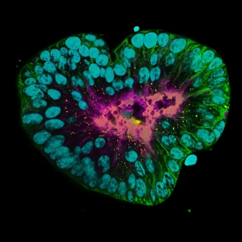
Banner image (above): Patient-derived pulmonary neuroendocrine tumor organoid labelled with DAPI (cyan), phalloidin (magenta), beta-catenin (green), and ZO-1 (yellow). Sample courtesy of Marina Cuenca and Heleen Jungen, Talya Dayton lab, EMBL Barcelona.
*This is a preliminary status of Viventis SCAPE. Specifications might be subject to modifications
Welcome and opening remarks
Binson John, Leica Microsystems

Micron at-a-glance: facilitating cutting edge research
Professor Lothar Schermelleh, Micron Oxford

Imaging approaches to visualize organoid complexity
Emmanuelle Steib, Leica Microsystems
Spatial biology as a tool to characterize retinal organoids
Dr Colin Chu, University College London, Institute of Ophthalmology
Visualizing life in 3D with innovative lightsheet solutions:
an introduction to Viventis SCAPE and Viventis Deep
Tom Phillips, Leica Microsystems