See the Hidden – Artificial Intelligence in Microscopy
Dr. Luciano Lucas
Director Leica Aivia Leica Microsystems
Read BioLuciano studied Biology at the University of South Wales (UK), where he focused on population genetics and carcinogenesis. He earned his PhD at the University of Nottingham (UK) with research work in the fields of vascular targeting therapies, intra-vital microscopy and image analysis. He then joined Bitplane/Andor, where he took care of Imaris’ roadmap, commercialization and customer support for over 4 years. In the last 5 years, Luciano has led the creation of Aivia (AI – Visualization – Image Analysis). Leica Microsystems acquired Aivia in March 2021.
Close
Dr. Peter Engerer
Product Manager Software Leica Microsystems
Read BioDr. Peter Engerer studied Biomedicine in Wuerzburg, Vienna, Singapore and Berlin. In 2016, he earned a Ph.D. from TU Muenchen for work on the development and application of cutting-edge in-vivo imaging of the nervous system. After consulting several years on software engineering and data science for the Engineering group, he joined Leica Microsystems in 2020 as a Global Product Manager for software.
Close
Dr. Patrice Mascalchi
EMEA Sales Application Specialist Aivia
Read BioPatrice comes from a biotechnology engineering background and holds a PhD in Cell Biology and Microscopy. He has strong skills in microscopy, image analysis and programming, acquired in the course of his work in various light microscopy core facilities in France and the UK. Patrice is now focusing on advanced image analysis including artificial intelligence.
CloseIntroduction to AI for Microscopists: all you need to know
Machine and deep learning technologies already play a major role in everyday life – think Alexa, Tesla’s Autopilot, Spotify, Amazon or YouTube. In the last 3 years, microscopists have started to use “AI based” solutions for image acquisition optimization (smart microscopy), object classification, image classification, segmentation, restoration, super resolution and virtual staining. In this talk we focus on the basic concepts of machine learning for microscopy applications and image analysis. We will discuss the key terminology often used by AI specialists that can be hard to decipher for non-experts in that space.
A powerful combination for live-cell imaging: STELLARIS confocal microscope platform meets artificial intelligence
Confocal imaging is the gold standard for high-resolution 3D imaging of live cells. While historically many innovations have focused on advances in hardware components, imaging improvements by software means are gaining more traction. I will give a preview to a novel denoising technique for live-cell experiments, which uniquely combines classical image processing with AI.
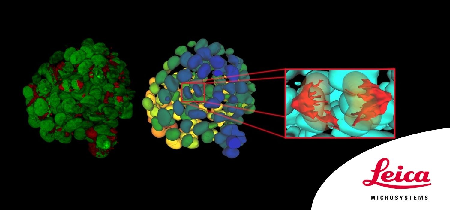
Aivia, the future of AI microscopy
Aivia is the most complete, AI-powered, image visualization and analysis platform available today.
With AI at its core, Aivia bridges the gap between biology and computer science to provide AI access to all and a radically simplified segmentation process to all. Using Aivia, biologists can easily extract insights from large and highly complex images in just minutes.
Aivia covers nearly all applications and is available locally or on the web, giving you total freedom on a single platform. Live demonstrations with the software will be performed during the workshop.