See the Hidden: Advancing Cryo Workflows for Cellular Discovery
Evgenia Zagoriy
Cryo-ET, Cryo-CLEM-FIB/SEM-TEM & cryo sample prep specialist, EMBL Heidelberg, Germany
Read BioEvgenia Zagoriy is an expert in cryo-FIB preparation of vitrified biological specimens, in situ cryo-electron tomography, and cryo-CLEM. She has been part of Julia Mahamid’s group at EMBL Heidelberg since 2017. Previously, during her PhD and postdoc at Max Planck Institutes, she studied chromosome biology and cell division. She holds a Master’s in Biochemistry from Aristotle University of Thessaloniki.
Close
Manon Demulder
Postdoctoral Researcher, Biozentrum Basel
Read BioManon is a structural biologist studying carbon fixation in algae using cryo-electron tomography. Her work combines mechanistic studies with environmental sampling workflows that extend cryo-ET to natural ecosystems. She completed her PhD at the Vrije Universiteit Brussel and is currently a postdoctoral researcher at the Biozentrum, University of Basel.
Close
Sofie van Dorst
PhD candidate in Structural Biology, Biozentrum Basel
Read BioSofie is a biologist and PhD student in the Cell Architecture group at the Biozentrum, University Basel. She studied at Utrecht University (NL) where she acquired a background in molecular and cellular biology. Her research centers around cyanobacteria and algae, she uses correlative light and electron microscopy to understand their photosynthesis from an ultrastructural perspective.
Close
Jan Groen
PostDoc | Microscopist | Cryo-Imaging, Pasteur
Read BioJohannes (Jan) Groen studied Biotechnology at Wageningen University (NL), where he developed a passion for microscopy. His PhD at the Mistral Beamline (Alba Synchrotron, Spain) focused on correlative light and X-ray microscopy to study therapeutic protein–nanomaterial interactions. As a postdoc at Institut Pasteur, he advanced cryo-CLEM workflows to study early SARS-CoV-2 infection. Now, at Synchrotron Soleil, he develops multimodal imaging workflows for complex biological samples.
Close
Vanessa Augustin
Advanced Workflow Specialist, Leica Microsystems
Read BioVanessa Augustin studied Molecular Cell Biology and Neurobiology at the Department of Animal Physiology in Kaiserslautern, later focusing on Human Biology and Human Genetics. Her research explored neuronal development, and the intracellular signaling pathways of the amyloid precursor protein, which is implicated in Alzheimer’s disease.
Close
Andreia Pinto
Advanced Workflow Specialist, Leica Microsystems
Read BioAndreia Pinto began her career as a Biomedical Scientist in Histocellular Pathology before focusing on Electron Microscopy. She spent over a decade managing an EM facility at a research institute in Lisbon, supporting projects from fundamental to translational research. After joining the Royal Brompton Hospital in 2019, she trained a deep learning platform (GETi) to detect ciliary defects in EM images for PCD diagnosis and led studies on SARS-CoV-2 infection of the respiratory airway. Now based at EMBL Heidelberg, she works with Leica Microsystems as a Workflow Application Specialist in sample preparation for electron and cryo-electron microscopy.
CloseLearn from the experts how to refine every step of your cryo workflow, from vitrification to imaging, to preserve native structure, minimise artefacts, and reveal the hidden details that drive cellular discovery.
These expert talks will show how you can:
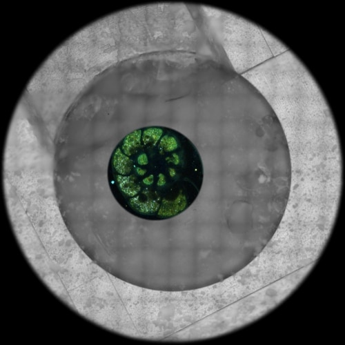
- Go beyond cryo-confocal: Use spectral signatures to unlock new layers of cellular information.
- Correlate across scales: Learn how to integrate cryo light and electron imaging for richer insights.
- Bridge modalities: Explore how X-ray tomography connects to cryo-EM analysis.
Cryo specimen preparation is transforming cell biology by preserving cellular structures with near-native fidelity. Achieving artifact-free samples, however, demands precise control at every stage.
In this See the Hidden event, leading experts from EMBL, the Pasteur Institute, Biozentrum Basel and Leica Microsystems reveal the workflows, tools, and techniques that make high-quality cryo imaging possible.
We’ll explore practical solutions for optimising instrumentation such as the STELLARIS Cryo, for maintaining cryogenic conditions across scales, and integrating complementary imaging modalities to extract more meaningful data from every sample.
This event will give you expert insight into how to push the limits of resolution and reveal the hidden details within your cells.