Multiscale Imaging Workflows for Organoid Imaging
Dr. Gustavo Quintas Glasner de Medeiros
Staff of Professorship for Multicellular Systems, ETH Zürich
Read BioGustavo completed his PhD at the European Molecular Biology Laboratory in Heidelberg, where he focused on deep-tissue light-sheet microscopy. He joined the Liberali Lab as a Postdoctoral Fellow in 2018 and became a Research Associate in 2022. Currently, as Staff of the Professorship for Multicellular Systems at ETH Zürich, he manages imaging workflows, microscopy development, and research data management. His research centers on developing imaging and perturbation methods to study the dynamics of symmetry-breaking during intestinal organoid growth.
CloseDiscover effective multiscale imaging workflows that integrate fixed and live microscopy to overcome the challenges of visualizing and quantifying organoid behavior.
In this webinar, you will:
• Discover techniques to extract and analyze multiscale data for meaningful biological interpretation
• Learn how to generate statistically robust organoid datasets with high-content imaging
• Gain strategies to perform and optimize long-term live imaging using light-sheet microscopy
• Understand the importance of collaborative, open-source tools for standardizing reproducible organoid imaging
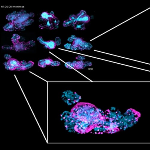
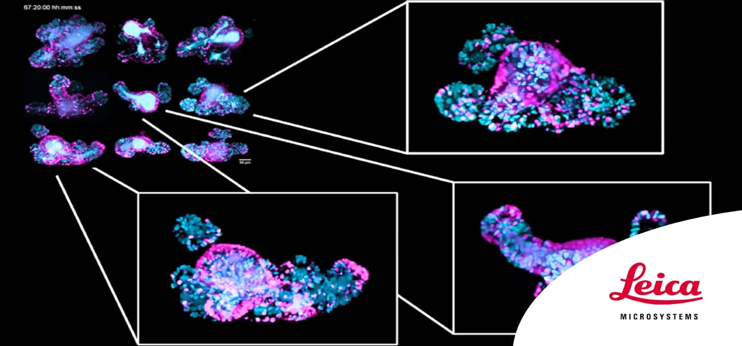
Organoids provide a powerful and physiologically relevant in vitro model to study tissue homeostasis, regeneration, and disease. However, their complex 3D architecture presents fundamental challenges for imaging—particularly in capturing both large-scale statistical information and dynamic biological processes.
In this webinar, you’ll learn how combining fixed-sample high-content imaging and live light-sheet microscopy delivers a complete picture of organoid biology—enabling phenotypic landscape profiling across large datasets while revealing the cellular mechanisms that drive organoid development. We’ll explore practical workflows for capturing and analyzing multiscale data and discuss how collaborative, open-source approaches are helping establish new standards in organoid imaging and data handling.
You will then discover how light-sheet microscopy has become the technique of choice for volumetric live imaging of 3D samples and gain insight into the key features of Viventis Deep ⎼ Leica’s open-top dual-view light-sheet microscope designed for long-term live imaging of multicellular samples.
Join us to understand how these imaging techniques can help you obtain reproducible, high-quality data and reveal deeper biological meaning from your organoid models.