Illuminating Nanomaterial–Cell Interactions by Confocal Microscopy

Barbara Rothen-Rutishauser
Professor of BioNanomaterials, Adolphe Merkle Institute, University of Fribourg
Read BioProf. Dr. Barbara Rothen-Rutishauser earned her PhD in cell biology from ETH Zurich in 1996. After positions at ETH Zurich and the University of Bern, she became co-chair of BioNanomaterials at the Adolphe Merkle Institute, University of Fribourg in 2011. Her research focuses on cell–nanomaterial interactions and the development of 3D epithelial tissue models. She is Associate Editor of Particle and Fibre Toxicology and has served on the SNSF Research Council since 2025.
Close
Luigi Di Stolfo
PhD student, Adolphe Merkle Institute, University of Fribourg
Read BioLuigi Di Stolfo is a PhD student in the BioNanomaterials Group at the Adolphe Merkle Institute. He uses confocal fluorescence microscopy and quantitative image analysis to study nanomaterial–cell interactions using in vitro cell models, focusing on how cellular variables such as spatial arrangement, morphology, and activation state modulate nanoparticle uptake and intracellular trafficking.
CloseSee how confocal microscopy reveals real-time nanomaterial uptake and intracellular trafficking in 3D, allowing you to study nano–bio interactions that shape diagnostics, targeted drug delivery, and safety outcomes.
In this webinar, you will learn how:
-
To design confocal imaging experiments that show where nanomaterials localize and how they move within cells
-
Nanomaterial properties influence cellular processing and how to interpret these effects in imaging studies
-
Different cell models and phenotypes affect nanomaterial internalization
-
A bioprinted epithelial gradient model reveals how cell density and tissue-like organization influence nanomaterial uptake
Engineered nanomaterials possess unique properties that are revolutionizing medicine through advanced diagnostics, targeted drug delivery, and tissue engineering. Their success depends on a detailed understanding of how they interact with single cells, which is relevant for efficacy and safety studies.
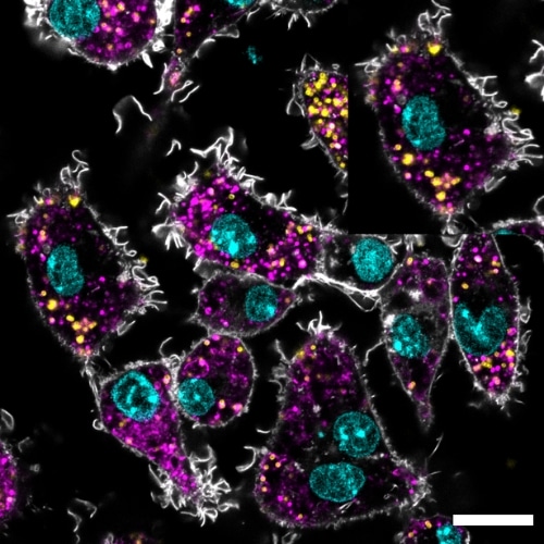
Confocal fluorescence microscopy offers powerful insight into these interactions by visualizing fluorescently labeled nanomaterials within complex cellular environments.
This webinar will highlight how confocal microscopy enables high-resolution 3D imaging and real-time analysis of nanomaterials uptake by cells and intracellular trafficking, particularly along the endolysosomal pathway. We will discuss how the physico-chemical properties of nanomaterials determine their cellular interactions, and how different cell models, such as epithelial barriers and immune cells, as well as altered phenotypes (e.g., inflamed or aging cells), influence nanomaterial uptake.
Special attention will be given to the long-term intracellular fate of nanomaterials and clearance mechanisms, critical parameters for medical applications.
The presentation will conclude with a recent study investigating how human lung epithelial cell density modulates nanomaterial uptake. Using a reproducible bioprinting drop-on-demand gradient approach, we examined relationships between cell density, accessible cell membrane, proliferation, and nanomaterial internalization by confocal microscopy, providing new insights into how tissue structures shape nano–bio interactions.