High-Resolution 3D Imaging Uncovers Vascular Control of Tissue Ageing

Dr. Anjali Kusumbe
Group Leader, Kennedy Institute of Rheumatology, Nuffield Department of Orthopaedics, Rheumatology and Musculoskeletal Sciences (NDORMS), University of Oxford
Read BioDr. Anjali Kusumbe is the head of the Tissue and Tumour Microenvironments Group at the Kennedy Institute of Rheumatology (University of Oxford).
She was awarded MRC Career Development Fellowship and ERC Starting Grant following her postdoctoral research at the Max Planck Institute for Molecular Biomedicine, Germany, on bone angiogenesis. She has received multiple awards, including the Werner-Risau Memorial Award and Iain T Boyle Award, for her work.
Webinar first broadcast Sep 15, 2021.
Ageing is associated with alterations in tissue homeostasis and a decline in tissue functions. Blood vessels provide supportive microenvironments for various cell types to maintain tissue homeostasis and organ function. However, ageing of the vasculature and how it contributes to changes in tissue physiology is poorly understood.
In this webinar, Dr. Kusumbe will demonstrate age-related changes in vascular microenvironments through single-cell resolution 3D imaging of young and aged organs. These changes include an age-dependent decline of capillary, artery, and pericyte numbers and increased fibroblast abundance in mice and humans, as revealed by tissue maps. Importantly, vascular attrition precedes the appearance of cellular hallmarks of ageing, such as senescence and mitochondrial dysfunction. Endothelial cell-specific genetic experiments confirmed that vascular perturbations drive cellular changes coupled with ageing, while genetic lineage tracing experiments, combined with imaging, revealed pericyte-to-fibroblast differentiation dictated by age-related molecular changes in the endothelium. Finally, pericyte-derived fibroblasts contributed to injury-induced organ fibrosis and experimental arthritis.
These findings unearth vascular attrition marked by pericyte-to-fibroblast differentiation, as a primary hallmark of ageing tissues.
To facilitate further research, Dr. Kusumbe’s group provides a freely available resource of 3D tissue maps with age-related features of vascular microenvironments.
In this webinar, you will discover:
- How tissue maps and genetic studies are providing new insights into the hallmarks of tissue ageing;
- How high-resolution imaging using the THUNDER Imager 3D Tissue is contributing to this research;
- How to access a freely available resource of 3D tissue maps with age-related features of vascular microenvironments.
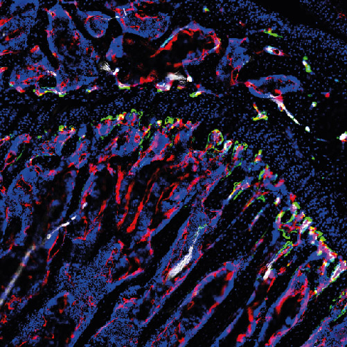
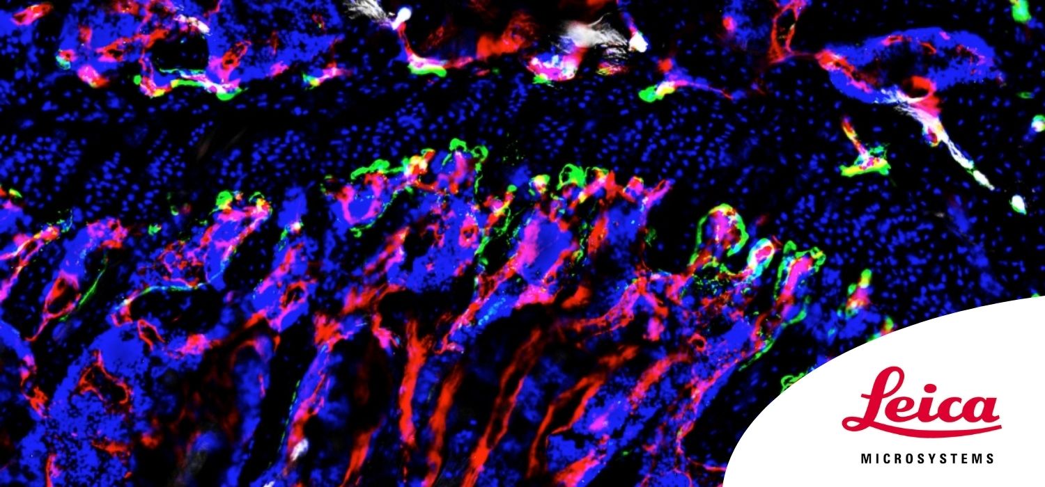
Image: Fluorescently stained mouse tibia, imaged with a THUNDER Imager 3D Tissue.
Courtesy of Dr. Anjali Kusumbe, Kennedy Institute of Rheumatology, NDORMS, University of Oxford, UK.