From Organs to Tissues to Cells: Analyzing 3D Specimens with Widefield Microscopy


Dr. Selina Keppler
Junior Group Leader Technische Universität München, Munich (TUM)
Read BioSelina did her PhD in Immunology, investigating the role of cytokines in T cell activation and memory formation during infection. Continuing her research on actin regulators and their role in B cell receptor signaling, Selina spent several years as Postdoc at the Francis Crick Institute London (former Cancer Research UK) in the lab of Facundo D. Batista. During her work in London, Selina first started imaging (confocal, 2-photon and TIRFM) and since then is hooked on colorful images. In late 2017, she started as Junior PI at the Center for Translational Cancer Research (TranslaTUM) in Munich. Selina and her team investigate the modulation of immune responses through the actin cytoskeleton during inflammation. In order to better understand the interplay between tissue niches, metabolism and immune cells, the Keppler lab also employs and advances imaging protocols. Recently, the Keppler lab implemented 3D imaging of organs or large tissue pieces in their workflow, thereby trying to push the borders of the THUNDER Imager a little.
Close
Dr. Falco Krüger
Advanced Workflow Specialist Widefield Life Science Research Division Leica Microsystems
Read BioAdvanced Workflow Specialist Widefield
Life Science Research Division
Falco did his PhD in Biology in the Plant Cell Biology group of Prof. Karin Schumacher at the Centre for Organismal Studies (COS) in Heidelberg, Germany. For his research on vacuole biogenesis in Arabidopsis thaliana, he used several different confocal microscopy techniques to understand the contribution of subcellular compartments to the development of one of the most prominent plant cell organelles.
By joining Leica Microsystems in May 2018, he switched gears, and started focusing on advanced widefield microscopy. Since the THUNDER launch earlier this year he has been hosting many workshops all over Germany, Austria and Switzerland, demonstrating the power of the new THUNDER Imagers to the scientific community. He enjoys getting to know different customers and their specimens, applications and imaging workflows.
In this webinar, you will discover:
- Different life science applications imaged with THUNDER Imagers;
- Quantitative imaging workflows of specimens of varying sizes and features;
- Cross-platform imaging workflows using different Leica microscope types;
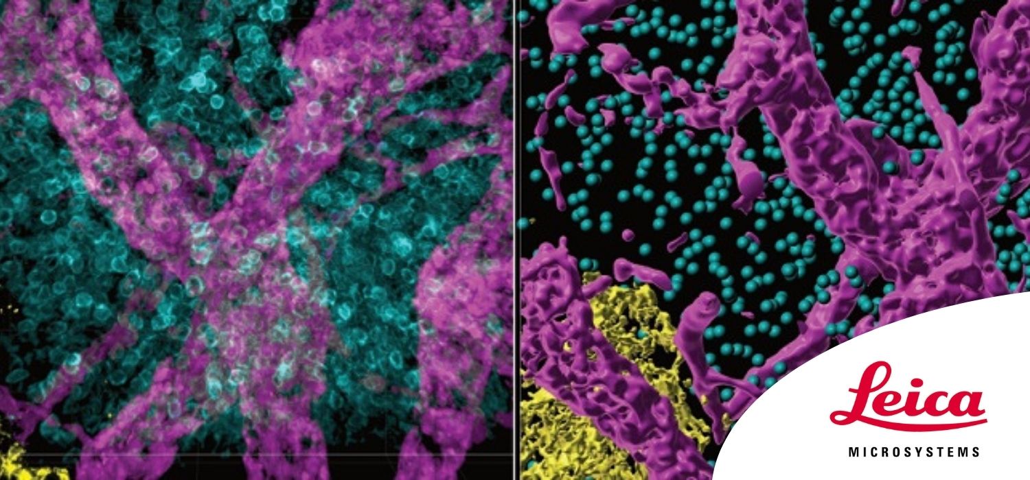
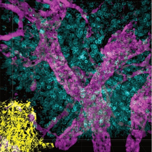
- An example of how THUNDER Imagers were used to perform quantitative single-cell analysis in cleared lymph nodes.
Abstract
It’s common in the life sciences to investigate a broad range of specimen types, from relatively flat mono-layer cell cultures to quite thick spheroids/organoids, tissue sections, or even whole model organisms.
Unfortunately, classic widefield microscopy is of limited use for life scientists investigating volumetric specimens as it can be challenging to get recorded data of sufficiently high quality. This is particularly true for the feature analysis in thick 3D samples because of the contribution of out-of-focus light.
However, this changed recently with the introduction of the Leica THUNDER Imager family.
In this webinar, you’ll discover the different life science applications that can be imaged with THUNDER Imagers, see the quantitative imaging workflows of specimens of varying sizes and features, and uncover the cross-platform imaging workflows using different Leica microscope types.
In addition, Dr. Selina Keppler, Junior PI at the Center for Translational Cancer Research (TranslaTUM) in Munich, gives us an exclusive insight of her work investigating the modulation of immune responses during inflammatory processes. For her research, the interaction of small cell populations within whole organs or tissue niches is of high importance. She discusses existing imaging workflows in her lab and how she pushed the limits of THUNDER Imagers to do quantitative single-cell analysis in cleared lymph nodes.
