Discover The Power of 3D High-Multiplex Imaging Across Scales

Dr Emmanuelle Steib
Advanced Workflow Specialist, Confocal Microscopy, Leica Microsystems
Read BioEmmanuelle currently works as an Advanced Workflow Specialist in Confocal Microscopy at Leica Microsystems. She was formerly an MSCA fellow at Imperial College London (UK), and has a PhD in Life Science from the University of Geneva (CH) and PharmD from the University of Strasbourg (FR).
CloseRecorded live from Imperial College London.
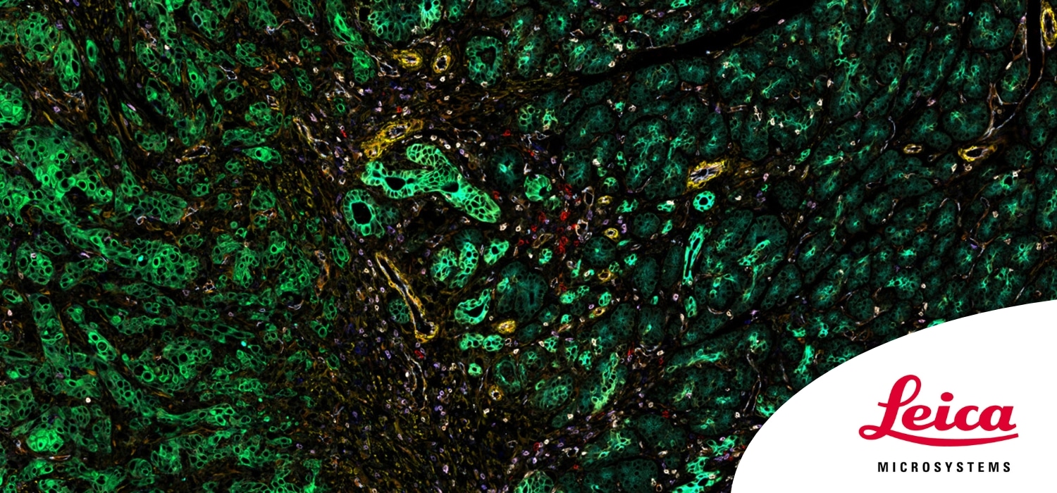
In this video, Dr Emmanuelle Steib presents the experimental design and considerations for high multiplexing imaging experiments with 15+ fluorophores on a single sample. Learn about appropriate fluorophore panels, sample preparation and the challenges and tools required for successful imaging. Emmanuelle shows how imaging high multiplexing targets on a single sample in one go (one round of labeling and imaging) can be exploited for 3D spatial omics analysis. Furthermore, we see how Aivia – Leica’s cutting-edge image analysis software – enables robust and efficient analysis of high multiplexing imaging data, providing researchers with powerful tools to extract meaningful insights from complex spatial biology experiments.
